What is a liver Shunt?
 A liver shunt is a blood vessel that carries blood around the liver instead of through it. In some animals a liver shunt is a birth defect (“congenital portosystemic shunt”). In others, multiple small shunts (“acquired portosystemic shunts”) form because of severe liver disease such as cirrhosis.
Why do congenital shunts develop?
All mammalian fetuses have a large shunt (“ductus venosus”) that carries blood quickly through the fetal liver to the heart. Since the mother’s liver does the work of filtering out toxins, storing sugar, and producing protein for her unborn babies, liver function is not needed in the fetus. This ductus venosus is supposed to close down shortly before or after birth as the baby’s liver begins to work. In some individuals the shunt doesn’t close down; it is then called a “Patent Ductus Venosus”, or an intrahepatic shunt. In other animals, a blood vessel outside of the liver develops abnormally and remains open after the ductus venosus closes. This is called a congenital extrahepatic shunt.
Why do animals with shunts have problems?
In the normal animal, food and other digested materials are broken down or digested in the intestines and absorbed into the portal blood stream, where they are carried to the liver. The liver stores some of the food for energy, processes some of it into safe chemicals and uses some of it to make proteins and other substances. Because the blood bypasses the liver in dogs with shunts, toxins may build up in the bloodstream or kidneys. Additionally, the animal lacks the necessary materials to give it a ready source of energy and to help it grow.
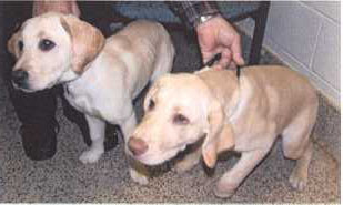
Guess Which Sibling Has The Shunt
What are the clinical signs of a liver shunt?
Clinical signs are often seen at a young age and include small stature, poor muscle development, behavioral abnormalities (circling, disorientation, unresponsiveness, staring into space, head pressing), seizures and quiet demeanor. Other less common signs include drinking or urinating too much, apparent blindness, diarrhea, and vomiting. In some animals the signs are associated with eating protein. Other animals are diagnosed when they take a long time recovering from anesthetics (i.e. barbiturates) or sedatives (i.e. acepromazine). Some animals show no signs until they are older, when they develop bladder and kidney infections and stones.
What breeds are commonly affected with shunts?
Small breed dogs tend to have shunts that form outside of the liver (“extrahepatic”). In the United States, Yorkshire terriers have almost a 36 times greater risk of developing shunts than all other breeds combined. Extrahepatic shunts can be seen in any small breed but are also reported commonly in Schnauzers, Maltese, Dachshunds, Jack Russell terriers, Shih Tzu, Lhasa Apso, Cairn terriers and Poodles. Large breed dogs tend to retain the fetal liver shunt (patent ductus venosus), or “intrahepatic” shunts. In the Netherlands, about 2% of the Irish Wolfhounds are born with intrahepatic shunts. Intrahepatic shunts can be seen in any large breed dog and have been reported in some small breed dogs (especially poodles); in the United States, we see them most often in Labrador retrievers. Australian shepherds, Australian cattle dogs, Samoyeds, and Old English sheepdogs are also commonly reported.
Are shunts hereditary?
A disease is likely to be hereditary if it occurs more commonly in one breed than others; if it occurs in a family of dogs; or if it or a closely related disease is proven hereditary in other breeds or species. Liver shunts are considered hereditary in Irish Wolfhounds, Cocker Spaniels, Maltese, and Yorkshire terriers, and are probably hereditary in several other breeds. The affected dog should be castrated or spayed and, because the mode of inheritance is not known, it is best to avoid breeding the parents.
How is a shunt diagnosed?
On blood work, dogs with congenital liver shunts usually have low blood urea nitrogen (BUN) and albumin concentrations. They may be slightly anemic or have red blood cells that are smaller than normal (“microcystosis”). They may also have increases in liver enzymes (“AST”,”ALT”). Their urine may be dilute or infected and contain small spiky crystals (“ammonium biurate”). None of these laboratory changes are specific for a liver shunt; however when veterinarians see these abnormalities, they will usually measure bile acid or ammonia concentrations to evaluate liver function. A liver shunt cannot be definitively diagnosed by blood work; shunting can only be found with advanced techniques such as scintigraphy, ultrasound, portography, Cat scan (“CT”), MRI, or exploratory surgery.
What are bile acids?
Bile acids are produced in the liver and stored in the gallbladder between meals. They are released into the intestines to help break down and absorb fats and are reabsorbed and stored again until they are needed. Dogs with liver shunts have increased blood bile acid concentrations because the liver does not get a chance to remove and store these chemicals after they are reabsorbed.
Do all dogs with shunts have high bile acids?
Dogs with shunts will almost always have high bile acids 2 hours after eating, and at least 95% of dogs will have high bile acids after a 12 hour fast. Samples are taken at both time periods (“fasting” or “preprandial”, and “fed” or “postprandial”) for several reasons. Some dogs normally release bile acid in the middle of the night and therefore naturally have a higher than normal fasting sample. Other dogs may have fat in their blood (“lipemia”) after eating, which can interfere with the test. If only one blood sample can be obtained, it is best to take it 2 hours after eating.
Do all dogs with high bile acids have shunts?
Bile acids can be increased with any liver disease. Bile acids can also be mildly increased in normal dogs, particularly in some breeds (such as Maltese) where chemicals in their blood interfere with the test. Most dogs with liver shunts have fed bile acids over 100 (normal <15-20). If the bile acids are only mildly increased or the animal seems normal, many veterinarians will simply rerun the test in 3-4 weeks.
What is scintigraphy?
Scintigraphy is a nuclear scan that measures blood flow. To evaluate a dog for a shunt, a radioactive material is inserted into the colon (by a high enema) and the animal is scanned with a special camera hooked to a computer. The computer measures the amount of radioactive blood in the heart and in the liver and compares the two. Normal animals have a shunt fraction (amount of blood in the heart divided by amount in the liver) of less than 15%. In other words, at least 85% of the radioactive material ends up in the liver. Dogs with shunts usually have shunt fractions >60%, because most of the blood bypasses the liver and goes straight to the heart. Scintigraphy is safe and quick but does require heavy sedation or anesthesia. Animals must be hospitalized for at least one night after the procedure until they have expelled the radioactive material by defecation and urination.
Scintigraphy tells us that shunting is present; however, in many cases the veterinarian cannot tell whether the shunt is congenital or acquired.
What is a portogram?
A portogram is an x-ray of the blood vessels to the liver. Because blood vessels are not easily seen on regular x-rays, a contrast material (a liquid that looks white on x-rays) must be injected into the blood vessel in the abdomen. The injection can be performed through a surgical incision into the belly; by injecting the spleen directly through the skin; or by passing a catheter down the jugular vein (in the neck), through the heart, and towards the abdomen. Portograms usually require anesthetic and are more invasive than scintigraphy. They are usually quite safe, however, and are able to provide a picture of the shunt so that the veterinarian can see where it is located and whether there is more than one.
Can a shunt be diagnosed with ultrasound?
Some veterinarians are able to find a shunt by ultrasounding the liver. Diagnosis of a shunt with ultrasound requires lots of experience and usually a specialized machine (“Doppler”) that can detect blood flow. Shunts, particularly those outside the liver, can be easily missed especially if the dog is small or wiggly, or the ultrasonographer is inexperienced.
Can a shunt be diagnosed with liver biopsy?
In animals with shunts, the liver is smaller than normal because it is atrophied from poor blood flow. On a liver biopsy, the tissue appears shrunken. Some of the vessels are very tiny, while others multiply in an attempt to improve blood supply and drainage. These changes are called hepatic microvascular dysplasia. Hepatic microvascular dysplasia (HMD or MVD) can also occur in dogs without liver shunts; therefore; other tests are needed to determine if a shunt is also present.
What medical management is needed for an animal with a shunt?
Dogs with shunts are usually stabilized with special diets and medications to reduce the amount of toxins that are produced and absorbed in the large intestines. Dogs that are severely ill may require intravenous fluids to restore blood sugar, an enema to remove intestinal toxins before they are absorbed, and medications such as valium to stop seizures.
Diet: Because many of the toxins produced in the intestines come from protein, it is important to reduce the amount of protein in the diet. Dog food for adults and puppies usually contains 25% and 29% protein respectively, and may have meat byproducts. Dogs with shunts need high quality proteins primarily made from milk or vegetable, and are restricted to a protein content of 18% or less (on a dry matter basis). The diets should be easily digestible, rich in antioxidants and vitamins, and low in copper and iron.
Lactulose: Much of the toxins absorbed from the intestines are produced by normal intestinal bacteria. Lactulose changes the pH in the large intestine, which decreases absorption of ammonia and other toxins and makes the environment unfavorable for the toxin-producing bacteria. It also encourages the intestinal contents to leave the area more quickly, so that toxins have less time to be absorbed. Lactulose is basically a sugar solution; its primary side effect is diarrhea. Because of this, veterinarians will instruct owners to adjust the dose so that the dog’s feces is soft but formed.
Antibiotics: If clinical signs are not controlled with a protein-restricted diet and Lactulose, veterinarians will often prescribe antibiotics to reduce the numbers of toxin-producing bacteria in the intestines. Antibiotics will also be needed if the animal has a urinary tract infection.
Can dogs with shunts be treated with only medical management?
Most animals improve immediately with proper diet and medicine, and about one third of the dogs treated medically will live a relatively long life. Unfortunately, over half of the dogs treated medically are euthanized, usually within 10 months of diagnosis, because of uncontrollable neurologic signs, such as seizures and behavior changes, or progressive liver damage. Dogs that tend to do well with long-term medical management are usually older at the time of diagnosis, have more normal blood work, and have less severe clinical signs. Surgery provides the best chance for a long healthy life in most dogs.
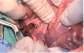
How is a shunt corrected surgically?
Because shunts inside the liver are more difficult to find and close off, surgery of dogs with intrahepatic shunts is best performed by a board certified surgeon (ACVS Diplomate). Surgery for congenital extrahepatic liver shunts is slightly easier, particularly if the veterinarian has a lot of experience, and is performed at most veterinary surgery referral centers. The surgeon must find the abnormal blood vessel and close it off to force blood flow back through the liver. Unfortunately, the blood vessels inside the livers of some dogs are so poorly developed that they will not open quickly. Therefore, most surgeons will use a devise that slowly closes the shunt such as the ameroid constrictor. Other options include placement of a suture or cellophane band around the shunt or coils inside the shunt. Placement of coils can be performed through a catheter in the neck (“jugular”) vein; however, because they tend to cause rapid obstruction of the shunt in animals, their use is still being researched.
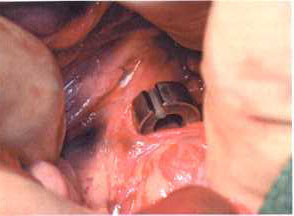
How does the ameroid constrictor work?
An ameriod constrictor is a metal band with an inner ring of casein, a protein found in milk. In the belly, the inner ring absorbs normal fluid and gradually swells, pressing on the shunt and encouraging it to scar shut. Shunts usually close within 3-4 weeks of ameroid constrictor placement. Because of the metal outer ring, the constrictor will always be visible on x-ray of the belly.
What are the complications of shunt surgery?
Surgery with the ameroid constrictor placement is faster and complications are few compared to other techniques, but the puppies can still get cold or develop low blood sugar during or after the procedure. Occasionally dogs will develop pain and bloating if the constrictor kinks the vessel or if a suture or a cellophane band is used. This can progress to shock and death, so animals must be watched carefully for several days after the procedure. A small percentage of dogs may also have seizures after surgery. Dogs with intrahepatic shunts are more likely to have complications and usually require several days of intensive care and possible blood transfusions.
What care is needed for dogs after shunt surgery?
Dogs are kept on a protein-restricted diet for at least 6-8 weeks after surgery. Lactulose can be continued as well, or can be gradually decreased over 2-4 weeks. Most dogs do not need antibiotics unless they have infections in the urine or other sites. The liver will begin to grow as the shunt closes, and will often be normal sized in 2-4 months. To check liver function, blood tests (BUN, albumin, liver enzymes and bile acids) are usually evaluated at 8-12 weeks after surgery. If these are still abnormal they are repeated in another 3 months. If they are normal, the diet is gradually switched to an adult maintenance dog food. A scintigraphy can be performed at 3-6 months to confirm the shunt is closed.
What is the prognosis for dogs after ameroid constrictor placement around a shunt?
Survival rate from the surgery is over 95% for dogs with shunts treated by ameroid constrictor placement and long-term prognosis is better with this technique than with most other methods. Many dogs are clinically normal within 4-8 weeks after the surgery. Long term, about 85% of dogs with liver shunts closed with ameroid constrictors do well clinically. About 15% continue to have problems, probably because the tiny blood vessels inside the liver were also abnormal. Usually these dogs develop multiple acquired shunts and must be managed with a protein restricted diet and Lactulose for life.
Dr. Karen Tobias
Karen Tobias, DVM, MS, Diplomate ACVS
Professor, Small Animal Surgery, University of Tennessee College of Veterinary Medicine
Regent, American College of Veterinary Surgeons;
President, Society of Veterinary Soft Tissue Surgery
University of Tennessee College of Veterinary Medicine
Univ phone (865-974-8387)
C247 Dept Vet. Clin. Sci
Knoxville TN 37996-4544
Tobias Office FAX 865-974-5554
Find a board certified surgeon near you at:
A Special THANK YOU to Dr. Karen Tobias for allowing me to use her brochure on my web site..
Click Below to open a PDF a copy of this page that you can save.